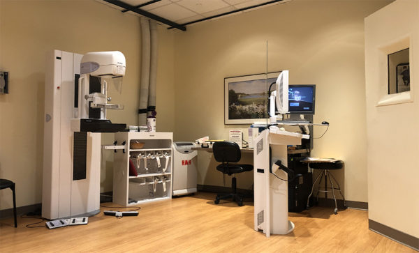
3-D Mammography uses advanced breast tomosynthesis technology, clinically proven to significantly improve the detection of invasive breast cancers, while simultaneously reducing the need to return for additional testing.

In conventional 2-D mammography, overlapping tissue is the leading reason why small breast cancers may be missed and normal tissue may appear abnormal, leading to unnecessary callbacks. A 3-D exam includes a three-dimensional method of imaging that can greatly reduce the tissue overlap effect.
During the tomosynthesis-dimensional portion of the exam, an X-ray arm sweeps in a slight arc over the breast, taking multiple images. A computer then converts the images into a stack of thin layers, allowing the radiologist to review the breast tissue one layer at a time. A 3-D exam requires no additional compression and takes just a few seconds longer than a conventional 2-D breast cancer screening exam.